


A team of Princeton University researchers recently elucidated the mechanisms involved in lung development using brown anole lizards as a model organism. Their developments represent a huge step towards the team’s continuing research surrounding lung tissue engineering and advancements.
In a study titled “Stress ball morphogenesis: How the lizard builds its lung,” published in the Dec. 22 issue of Science Advances, the research team outlines how the lizard breathes with elegant simplicity. Among biology’s most common themes is that of form and function: that the structure of an organism dictates its intended purpose. Morphogenesis is the process that causes the shape of a tissue or an organ to develop to serve its function. While scientists have known about the function of the lungs of lizards, the biological and physical basis of its morphogenesis have never been studied in detail.
In their effort to better understand morphogenesis, the team of researchers traveled to Florida to catch the brown anole lizards and create a suitable habitat system for the lizards to survive in Princeton. The team chose the brown anole lizard to focus on since the organism develops its lungs at a mind-blowingly fast rate — over a two-day period. Another major reason for the selection of the lizard was its simple lungs; they are composed of only two bags that have a bumpy surface, where oxygen exchange happens.
The developing lizard lung starts as a single hollow tube and inflates to a rounded balloon-like shape. As the expansion is happening, a stiff tissue called smooth muscle wraps around the outside of the big balloon and forms a barrier. As a result, once the balloon is expanding outward, it encounters a “mesh” of smooth muscle. The balloon then grows through the gaps in that mesh because it cannot grow where the mesh itself is. That balloon forms the unique corrugated structure of the lizard lung.
This knowledge has significant transferability to humans. In fact, it lays the framework for artificial lung synthesis by mirroring an already existing natural phenomenon.
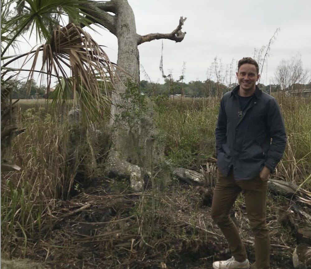
“My team is really interested in trying to come up with ways to build lungs for tissue engineering purposes,” said Celeste Nelson, the Wilke Family Professor in Bioengineering and the study’s principal investigator.
Her fascination with lungs is rooted in a personal story from her childhood.
“My grandfather only had one lung,” Nelson explained. “He lost a lung after serving in the military.”

As a kid, Nelson said she found it strange how her grandfather could still survive with one lung while not being able to do more intense physical activities, like running around.
When graduate student Michael Palmer GS ’21 joined Nelson’s lab, he had a strong interest in evolutionary biology and differences in tissue structure across different classes of vertebrates. His focus: chicken and lizard lungs.
“I am interested in studying diverse lung structures in order to better understand what’s possible within the field of lung development,” said Palmer, who earned his Ph.D. in Chemical and Biological Engineering.
The team deviated from convention in their approach, inspired by the evolutionary similarities across different species, especially as it pertains to the structure of the lung.
In addition to outlining the process of lung development in lizards, the research team also tested the physical nature of the process using a computational model. The model was developed in collaboration with the lab of Andrej Košmrlj, Assistant Professor in the Department of Mechanical and Aerospace Engineering.
In the end, using their findings of how the lizard forms its lung and the computational model that they had developed, the team was able to produce a model of the lizard lung system that works outside of the living organism. To model the balloon-like structure, they used Ecoflex, thin silicone elastomer sheets which are commonly used in the film industry. The team was challenged to find a material with roughly the same mechanical properties as the developing lizard lung.

Once the team succeeded at overcoming this challenge, they then 3D-printed muscle cells on top of that structure in a mesh pattern, similar to what smooth muscle looks like in the developing lung.
And there it was: a physical model of the lung similar to the one found in the living anole lizard.
“What I think is the most exciting part about this project is just that it combines so many different aspects of biology and engineering,” said Palmer. “On the one side, you have the pure developmental biology trying to understand how the structure forms and then we coupled that with computer-based simulations to better understand the physics. Finally, we used bioengineering to create a system that recapitulates the biology, but in a laboratory setting.”
“So many fields intersecting,” Palmer added.
Nelson reflected on the increased relevancy of the team’s work amid the pandemic.
“Since we’re in the middle of a pandemic, I think most of us are paying more attention to our lungs now. Before, they were rather unappreciated, despite being these fascinating structures,” said Nelson.
Her lab’s ultimate goal, which Nelson anticipates will probably take 20 or more years, is to build a fully-functioning lung inspired by nature.
Mahya Fazel-Zarandi is a staff writer for the ‘Prince.’ She can be reached by email at mahyaf@princeton.edu or on Twitter @MahyaFazel.
Read More

Undergraduate cyclist struck by University employee sues for damages
Haeon LeeThe student, who was seriously injured, is suing the University, the employee, and the owner of the vehicle for damages.
The student, who was seriously injured, is suing the University, the employee, and the owner of the vehicle for damages.

Former Princeton standout Xaivian Lee signs with the Cleveland Cavaliers on Exhibit 10 contract
Jordan HalagaoFormer Princeton men’s basketball player Xaivian Lee signed an Exhibit 10 contract with the Cleveland Cavaliers to participate in training camp and Summer League.
Former men’s basketball player Xaivian Lee signed an Exhibit 10 contract with the Cleveland Cavaliers to participate in training camp and Summer League.

Princeton names eight new trustees, increasing total trustee count by three
Nico David-FoxThe board now has 40 members, the maximum number permitted under its bylaws.
The board now has 40 members, the maximum number permitted under its bylaws.